Renal Regulation of Fluid and Electrolyte Balance

Comprehensive exploration of kidney physiology and the mechanisms governing water and electrolyte homeostasis.

Kidney anatomy and nephron structure

Overview of Renal Function

The kidneys are sophisticated organs responsible for maintaining fluid volume, electrolyte concentrations, and acid-base balance. Despite comprising only 0.5% of body weight, the kidneys receive approximately 20% of cardiac output, emphasizing their critical role in homeostatic regulation.

Fundamental Processes

Renal function operates through three primary mechanisms: glomerular filtration, tubular reabsorption, and tubular secretion. Glomerular filtration passively filters plasma across the glomerular barrier, producing approximately 180 liters of filtrate daily. The vast majority is reabsorbed; normally only 1-2 liters remain as urine. Selective reabsorption of essential substances in the renal tubules and collecting ducts recovers water, electrolytes, glucose, and amino acids while retaining waste products for excretion.

Glomerular Filtration

Glomerular filtration rate (GFR) depends on the balance between hydrostatic and oncotic pressures across the glomerular barrier. Hydrostatic pressure in the glomerular capillaries favors filtration, while plasma protein oncotic pressure opposes filtration. The glomerular barrier selectively permits passage of small molecules (water, glucose, electrolytes, urea) while restricting large molecules (proteins, blood cells).

Filtration Dynamics

Autoregulation maintains relatively constant GFR despite blood pressure fluctuations between 80-180 mmHg. When blood pressure increases, afferent arteriole vasoconstriction reduces glomerular capillary pressure. Conversely, decreasing blood pressure triggers afferent arteriole vasodilation, maintaining filtration pressure. This autoregulatory mechanism ensures stable filtration and prevents excessive fluid loss during postural changes or minor blood pressure variations.

Renal perfusion pressure reduction activates the renin-angiotensin-aldosterone system (RAAS). Juxtaglomerular cells in the afferent arteriole detect decreased pressure and release renin, initiating a cascade that ultimately increases angiotensin II production. Angiotensin II acts as a potent vasoconstrictor, increasing filtration pressure while promoting sodium and water reabsorption, thereby restoring blood volume and pressure.

Tubular Reabsorption and Secretion

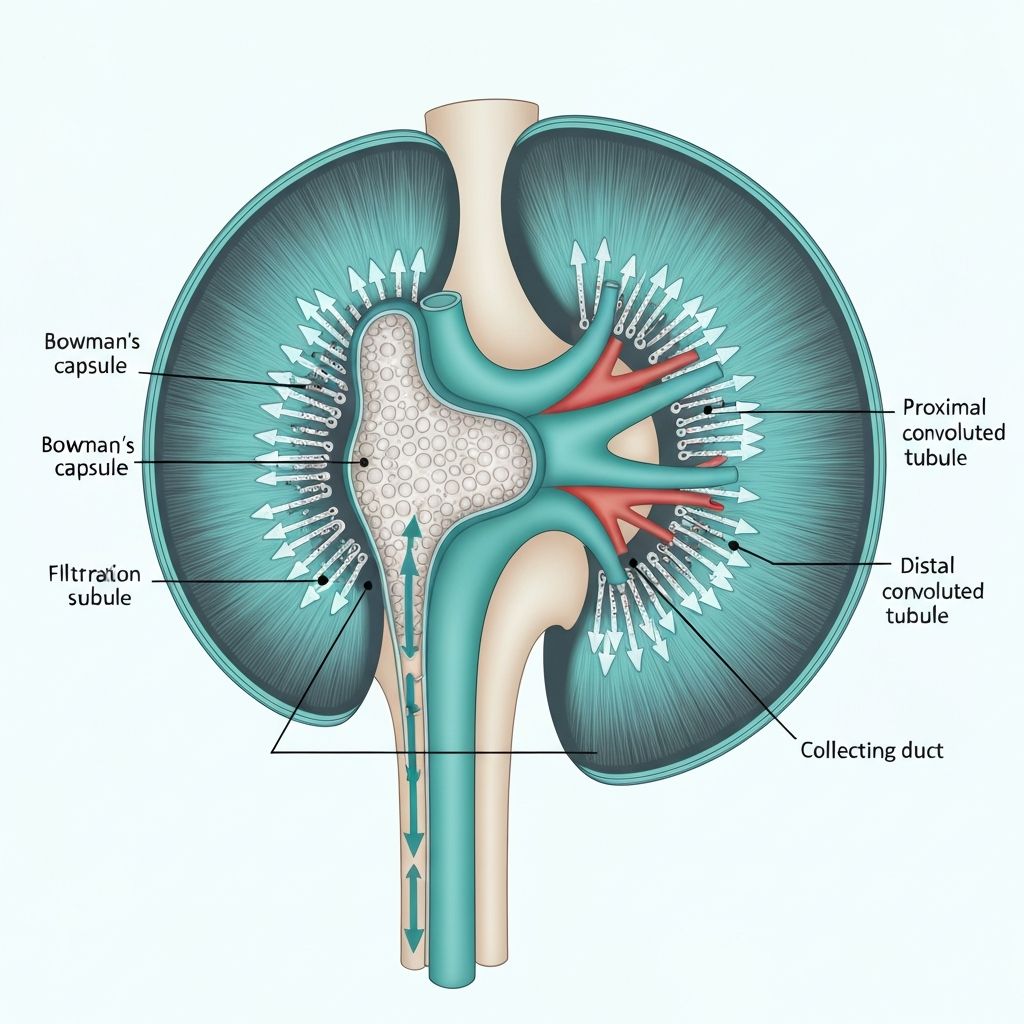
The renal tubules and collecting ducts are the primary sites of selective solute and water reabsorption. Different tubular segments possess distinct epithelial characteristics and transport capabilities, allowing fine-tuned control of substance recovery.

Proximal Convoluted Tubule

The proximal tubule reabsorbs approximately 65% of filtered sodium, chloride, glucose, amino acids, and water. Active transport via the sodium-potassium pump drives these reabsorption processes. Aquaporin-1 water channels facilitate obligatory water reabsorption through osmotic gradients established by solute reabsorption. This segment operates iso-osmotically; reabsorbed fluid osmolarity remains equal to plasma osmolarity.

Loop of Henle

The loop of Henle consists of descending and ascending limbs with distinct permeability properties. The thin descending limb is highly permeable to water but impermeable to solutes, allowing passive water reabsorption as filtrate moves toward the hairpin turn. The ascending limb is impermeable to water but actively reabsorbs sodium and chloride through active transport and cotransport mechanisms. This countercurrent mechanism establishes an osmotic gradient, concentrated in the medulla, that drives water reabsorption in collecting ducts.

Distal Convoluted Tubule and Collecting Duct

The distal tubule and collecting duct are primary sites of fine-tuned sodium and water reabsorption regulated by hormones. Aldosterone increases epithelial sodium channel expression and sodium-potassium pump activity, promoting sodium reabsorption and water reabsorption through osmotic coupling. Antidiuretic hormone (ADH) increases aquaporin-2 water channel expression in collecting duct principal cells, permitting water reabsorption in response to plasma osmolarity.

Detailed nephron structure and reabsorption mechanisms

Hormonal Regulation of Water Balance

Antidiuretic hormone (ADH), also called vasopressin, is synthesized in the hypothalamus and released from the posterior pituitary. ADH responds to changes in plasma osmolarity and blood volume, modulating water reabsorption in the collecting duct. Elevated plasma osmolarity stimulates ADH release, increasing aquaporin-2 expression and enabling concentrated urine production. Conversely, decreased osmolarity suppresses ADH, reducing water reabsorption and producing dilute urine.

ADH Mechanism of Action

ADH binds to V2 receptors on collecting duct cells, triggering a cascade that increases cyclic AMP. This second messenger activates protein kinase A, phosphorylating regulatory proteins that insert aquaporin-2 water channels into the apical membrane. This insertion increases membrane water permeability, allowing water to follow osmotic gradients established by solute reabsorption. When ADH levels decline, aquaporin-2 channels are internalized through endocytosis, reducing water permeability. This rapid responsiveness enables precise water balance adjustment.

Sodium-Potassium Regulation

Sodium and potassium distribution fundamentally determines cellular and systemic fluid balance. The kidneys excrete excess sodium while conserving potassium through selective tubular reabsorption and secretion.

Sodium Handling

Approximately 65% of filtered sodium is reabsorbed in the proximal tubule through active transport. An additional 25% is reabsorbed in the loop of Henle, particularly the thick ascending limb through the Na-K-2Cl cotransporter. The distal tubule and collecting duct reabsorb the remaining sodium through epithelial sodium channels (ENaC) regulated by aldosterone. This multi-segment reabsorption permits fine-tuned control of sodium excretion in response to dietary intake and hemodynamic changes.

Potassium Handling

All filtered potassium is reabsorbed in the proximal tubule and loop of Henle. Distal tubule and collecting duct potassium secretion regulates urinary potassium excretion. In the collecting duct, principal cells secrete potassium through ROMK channels and BK channels. Aldosterone increases principal cell potassium secretion by stimulating Na-K-ATPase expression and increasing epithelial sodium channel activity, which hyperpolarizes the membrane potential and facilitates potassium efflux. This aldosterone-dependent mechanism ensures potassium excretion matches dietary intake, preventing both deficiency and excess.

Acid-Base Balance Regulation

The kidneys contribute to acid-base balance primarily through bicarbonate reabsorption and hydrogen ion secretion. The proximal tubule reabsorbs approximately 90% of filtered bicarbonate through hydrogen ion secretion coupled with carbonic acid formation and dehydration to carbon dioxide, which diffuses into tubular cells.

Metabolic acidosis increases hydrogen ion secretion in the proximal and collecting ducts, promoting bicarbonate reabsorption. Alpha-intercalated cells in the collecting duct secrete hydrogen ions through H-ATPase pumps, allowing bicarbonate reabsorption from glomerular filtrate. Metabolic alkalosis reduces hydrogen ion secretion, permitting bicarbonate excretion in urine. The kidneys respond more slowly than respiratory buffering but provide essential long-term acid-base regulation.

Clinical Perspectives on Renal Function

Understanding normal renal physiology provides context for recognizing how various conditions affect fluid and electrolyte balance. Acute kidney injury, chronic kidney disease, and specific tubular disorders disrupt these regulatory mechanisms, potentially contributing to fluid retention, electrolyte imbalances, and altered acid-base status.

Certain medications affect renal function through various mechanisms. Loop diuretics inhibit the Na-K-2Cl cotransporter in the thick ascending limb, preventing sodium and chloride reabsorption and reducing the osmotic gradient driving water reabsorption. NSAIDs reduce renal blood flow by blocking prostaglandin-mediated afferent arteriole vasodilation, decreasing GFR. ACE inhibitors and ARBs dilate the efferent arteriole, reducing glomerular filtration pressure and GFR, particularly significant in individuals with underlying kidney disease.

Key Concepts Summary

  • Glomerular filtration produces approximately 180 liters of filtrate daily; autoregulation maintains stable GFR despite blood pressure fluctuations.
  • Selective reabsorption in proximal tubule recovers 65% of filtered sodium, glucose, amino acids, and water.
  • Countercurrent multiplication in the loop of Henle establishes osmotic gradients enabling water reabsorption.
  • ADH and aldosterone provide fine-tuned control of water and sodium reabsorption in the distal tubule and collecting duct.
  • Acid-base regulation maintains pH through bicarbonate reabsorption and hydrogen ion secretion.

Return to Blog